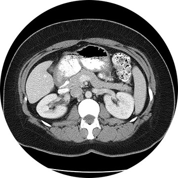
What is a CT scan?
A CT (Computed Tomography) scan is an advanced type of X-ray exam. Multiple X-rays are taken rapidly from a number of different angles around the body and then arranged by a high-speed computer to produce a cross-sectional view. CT may be used to visualize internal organs, head, neck, spine or extremities.
For some CT scans, the radiologist injects intravenous contrast medium or dye to highlight certain tissues for closer examination. Certain patients may also be required to drink oral contrast as well. A CT scan helps differentiate between healthy and diseased tissue, making it possible to accurately diagnose many diseases in their early stages.
When is a CT scan used?
CT scanning is generally used when your doctor needs more detailed diagnostic information than is possible from regular X-ray studies.
What happens during a CT scan procedure?
You will be positioned onto the table for the scan. You will feel the table move after each scan and may hear a whirring noise or high-pitched beep.
To get the most precise results, the technologist may ask you to hold your breath for a short time. Lie as still as possible to avoid blurring the images. You will be able to communicate with the technologist at all times during your scan. Actual time to acquire the images is often less than two minutes, but you should expect to be in the imaging room for approximately 10 to 15 minutes in its entirety.
You may leave immediately after your CT scan. If contrast was used, drink plenty of fluids, especially water, for the next 24 hours to help flush the contrast medium from your body. The radiologist will review your scans and send the results to your physician. Urgent findings will be called or faxed in to your physician very shortly after completion of your study.
What are the benefits and risks of a CT scan?
CT scans are among the safest exams we do. Your body will be exposed to a very small amount of radiation. If you are pregnant, you should not have a CT scan without first discussing the risks with your doctor. There is a small risk you will have an allergic reaction to contrast dye. Common minor allergic reactions may include hives and itching. More serious contrast reactions are very rare. Be sure to tell your health care provider if you know you are allergic to any medications or chemicals such as iodine. Our staff and physicians are trained and prepared to handle immediately any allergic reaction you might have and prevent any future occurrences.
Some CT Imaging Procedures Include:
The high-resolution medical images in 3D show a remarkable real-life appearance of your internal organs. The technology is far advanced over traditional CT imaging and provides excellent diagnostic information. Whole body scanning, coronary artery calcium scoring, virtual colonoscopy, lung screening, heart scanning—these are the advanced 3D imaging procedures available to patients at our facilities.
An angiogram is an X-ray exam of the arteries and veins to diagnose blockages and other blood vessel problems. It can reveal the integrity of the cardiovascular system in specific areas throughout the body. Combined with the use of intravenous contrast medium injected via a catheter, an angiogram identifies areas of blockage or damaged vessels within the circulatory system. CT and MRI may also be used to gain additional images of the arteries.
This exam is part of a sophisticated high-speed CT exam of the heart. During the scan, which takes just seconds, the equipment measures the amount of calcium present and calculates a score. The lower the score, the lower the potential risk of an adverse future cardiac injury. (Calcium often covers the atherosclerotic plaque that builds up inside arteries. This plaque and calcium can lead to narrowing of the inside of the arteries which could in turn lead to an increased risk of angina, and a heart attack.) This test can assess coronary heart disease, which is often asymptomatic and is the most common cause of death for patients in the United States.
This is an accurate and noninvasive imaging procedure used to assess and evaluate certain gastrointestinal problems, such as inflammatory bowel disease (including Crohn’s Disease), infectious enteritis, lymphoma or tuberculosis. It also can be used in patients with gastrointestinal bleeding to determine if a small bowel polyp is causing the bleeding. Enterography may be performed using MRI or CT.
A lung scan helps clinicians examine your lungs using a radionuclide and X-rays. A radionuclide is injected, and a camera can image how that liquid circulates in your bloodstream, specifically the supply to your lungs. You might also be asked to breathe a small amount of radionuclide mixed with oxygen through a face mask. The camera records where the air is going inside your lungs.

