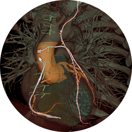
Chest imaging procedures are typically designed to assess cancer, toxin exposure, pulmonary abnormalities, embolism, inflammation, bronchial blockage, pleural effusion, angina, and other disorders.
Some Chest Imaging Procedures Include:
A chest X-ray is a very common medical procedure that can reveal the cause of chest pain, persistent cough or difficulty breathing. A very small dose of radiation is used to image the lungs, heart and other structures in the chest.
A lung scan helps clinicians examine your lungs using a radionuclide and X-rays. A radionuclide is injected, and a camera can image how that liquid circulates in your bloodstream, specifically the supply to your lungs. You might also be asked to breathe a small amount of radionuclide mixed with oxygen through a face mask. The camera records where the air is going inside your lungs.
If an abscess has formed in the lungs, an interventional radiologist can use images to direct placement of a needle into the affected area for drainage. The drainage tube (catheter) can be guided by CT, fluoroscopy, ultrasound or X-ray.


