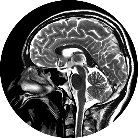
Our board-certified neuroradiologists investigate pathologies and injuries of the head, neck, and spine using the most advanced imaging technologies available. Using mainly MRI and CT, neuroradiologists diagnose abnormalities of the central and peripheral nervous system.
Some Brain & Spine Imaging Procedures Include:
An angiogram is an X-ray exam of the arteries and veins to diagnose blockages and other blood vessel problems. It can reveal the integrity of the cardiovascular system in specific areas throughout the body. Combined with the use of intravenous contrast medium injected via a catheter, an angiogram identifies areas of blockage or damaged vessels within the circulatory system. CT and MRI may also be used to gain additional images of the arteries.
Myelography is an exam in which contrast material is injected into your spinal column and then that contrast and spinal anatomy is imaged with CT technique. This allows the neuroradiologists to evaluate areas of nerve root impingement, canal narrowing, or disc protrusions. It is typically used when a patient is not a candidate for MRI.
NeuroQuant (NQ) is an artificial intelligence (AI) tool that calculates the volume of different substructures of the brain and compares those to a large normative age- and gender-matched database to determine whether the degree of brain volume loss is statistically significant for patient age. This can be used to improve the early detection of Alzheimer’s Disease (AD) or other neurodementia syndromes. NeuroQuant-MS is used to calculate the volume, number, and location of plaques in patients with multiple sclerosis. The software highlights new, enlarging, or shrinking plaques. This allows for accurate tracking of disease status over time. NQ can also be used to detect the location of a seizure focus in patients with epilepsy. It is also used in brain trauma patients or to assess brain development. Neuro-Quant has both MRI and CT applications. Other NeuroQuant-based AI tools in development include volumetric quantification and characterization of brain tumors.
We provide a range of interventional procedures for the management of spinal pain. They include:
Vertebroplasty and kyphoplasty are procedures that treat spinal fractures or compressed / collapsed vertebrae, often performed by a neuroradiologist. Vertebroplasty is the injection of a cement-like material into the bone to make it more stable. In kyphoplasty, the doctor first creates space by inflating a balloon-like device in the bone. The space is then filled with the cement material.
Discography is a procedure used to confirm that an abnormal disc is the culprit of pain. It is often used prior to a more invasive surgical procedure, to gather more information before that next step is taken. A contrast agent is introduced, and after the procedure a CT scan identifies leakage from the discs to identify any spinal disc herniation.
Facet and SI joint injections are used to alleviate joint pain, such as with arthritis or injury, or to determine the exact cause of pain. Under X-ray guidance, a clinician injects anesthetic directly into the joint or anesthetizes the nerves to carry pain signals away from the joint.
Nerve root injections are used to help ease back and leg pain when a nerve is inflamed or compressed as it passes from the spinal column between the vertebrae. The injection contains a steroid medication or anesthetic and is administered to the affected part of the nerve.
Epidurals can be diagnostic (an isolated nerve is injected with anesthetic to see if it is the source of pain) or therapeutic (both an anesthetic and an anti-inflammatory medication, such as a steroid (cortisone), are injected near the affected nerve).

