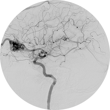
What is angiography?
Angiography is an X-ray exam of the arteries and veins to diagnose blockages and other blood vessel problems. It can reveal the integrity of the cardiovascular system in specific areas throughout the body. Combined with the use of some type of contrast medium injected via a catheter, the angiogram identifies areas of blockage or damaged vessels within the circulatory system.
When is it used?
Typically an angiogram serves to reveal whether there is a blockage or narrowing in a blood vessel that may interfere with the normal flow of blood through the body. The most common type of angiogram is the coronary angiogram, which identifies abnormalities in the blood circulation to and through the heart chambers. Other types of angiograms include cerebral (brain), extremity (arm or leg), renal (kidney), pulmonary (lungs), lymph vessels, and retinal (eyes). An angiogram could be used to discover aneurysms (an area of a blood vessel that bulges or balloons out), cerebral vascular disease (such as stroke or bleeding in the brain), or blood vessel malformations. Each of these requires the injection of some type of contrast in order to adequately view the blood circulation.
What happens during the procedure?
In the standard procedure, which is performed by an interventional radiologist, the doctor inserts a thin tube (catheter) into the artery through a small nick in the skin about the size of the tip of a pencil. A contrast agent (X-ray dye) is then injected to make the blood vessels visible on the X-ray. You will be awake during the procedure so that you can follow instructions, and your heart and blood pressure will be monitored. In many cases, the interventional radiologist can treat a blocked blood vessel without surgery at the same time the angiogram is performed. Interventional radiologists treat blockages with techniques called angioplasty and thrombolysis.
We can perform an initial, non-invasive evaluation of any part of the circulatory system; discuss this option with your doctor. For example, a CT angiography examines the blood vessels with the use of computerized tomography (CT), following the injection of a radiopaque contrast substance. Computerized tomography angiography (CTA) is a safe outpatient alternative to the standard catheter angiography.
What are the benefits and risks?
Major complications are rare. As with any procedure on the heart or blood vessels, an angiogram presents a risk of heart attack, stroke or infection.
Some Angiography Imaging Procedures Include:
An angiogram is an X-ray exam of the arteries and veins to diagnose blockages and other blood vessel problems. It can reveal the integrity of the cardiovascular system in specific areas throughout the body. Combined with the use of intravenous contrast medium injected via a catheter, an angiogram identifies areas of blockage or damaged vessels within the circulatory system. CT and MRI may also be used to gain additional images of the arteries.


