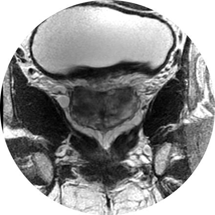
Prostate imaging centers on three major health problems of the prostate gland: cancer, benign enlargement, and inflammation (the cause of most urinary tract infections). Radiology plays an important role in diagnosing and treating these problems.
Some Prostate Imaging Procedures Include:
Magnetic resonance imaging (MRI) of the prostate uses a powerful magnetic field, radio waves and a computer to produce detailed pictures of the structures within a man’s prostate gland. It is primarily used to evaluate the extent of prostate cancer and determine whether it has spread. It also may be used to help diagnose infection, benign prostatic hyperplasia (BPH) or congenital abnormalities.
Treatment for prostate cancer can involve image-guided radiation therapy. In some advanced treatments, daily image guidance is increasingly used to improve the setup due to organ movement. Since the prostate position varies day-to-day depending on bladder and rectal filling, the prostate position must be localized and verified prior to each treatment.
In one method, several fiducial markers, or tiny pieces of biologically inert metal such as gold, are placed in the prostate gland before the simulation. Digital X-ray images are taken which localize the metallic markers to check the position of the prostate on a daily basis just before the treatment, and the prostate is appropriately adjusted and aligned to the planned high-dose radiation treatment field.
Another method involves using ultrasound to localize the prostate before each treatment. The patient is asked to keep his bladder full as much as possible in order to produce a good ultrasound image, and also to displace the bladder mucosa out of the radiation treatment field.
A third method involves the use of a low-dose computed tomography (CT) scan of the prostate area in the treatment couch right before each treatment to verify prostate position. Depending on your case and available technology at your treatment center, your physician will inform you of the type of image-guided radiation therapy you will receive.
Ultrasound is a non-invasive procedure that uses sound waves to image the structure and movement of the body’s internal organs, as well as blood flowing through blood vessels, in real-time. The prostate gland and surrounding tissue are examined by the insertion of an ultrasound probe into the patient’s rectum. There are no harmful effects, and it gives a clearer picture of soft tissues than X-ray images.


