
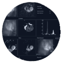
CARDIAC IMAGING
in the New York Metro
Every heartbeat tells a story. At Lenox Hill Radiology, we believe in the importance of capturing every detail to provide life-saving insights. Our leading-edge cardiac imaging solutions empower patients and physicians with the precision they need to understand, diagnose and treat heart conditions with confidence.
.
Lenox Hill Radiology's Comprehensive Cardiac Imaging Program is the New York Metro region’s most robust, non-invasive, outpatient, cardiac imaging program.
Our Cardiac Services
Cost Advantage
From our unmatched fleet of advanced, Low-Dose Cardiac CT units and dozens of upgraded Wide-Open Cardiac MRIs to our highly trained clinical and support staff, our Comprehensive Cardiac Imaging Program offers New York cardiology patients only the very best in diagnosis.
As an independent medical imaging provider operating outside of hospital affiliations, Lenox Hill Radiology offers affordable, high-quality, non-invasive cardiac imaging at a cost that is up to 50% lower than that of hospitals.
Safety & Comfort is our Top Priority
Early detection can lead to additional treatment options and a more favorable outcome. Our cardiac imaging program can provide a comprehensive evaluation of the heart, including its structure, function, blood flow, and tissue perfusion.
Our Cardiac CT exams are performed with low-dose protocols on the most advanced units. A Cardiac MRI at LHR provides a complete understanding of a patient's condition while also guiding treatment decisions. Advanced imaging can help a cardiologist tailor a treatment plan to each patient's unique needs, based on their individual cardiac anatomy and function.
Accurate Diagnosis
Advanced cardiac imaging techniques, such as Coronary Computed Tomography Angiography (CCTA), provide several advantages, including a more accurate diagnosis.
Our Comprehensive Cardiac Imaging Program employs the most advanced cardiac imaging techniques and tools available in order to provide the highest quality images of the heart structures and morphology, as well as the newest platforms and tools that help demonstrate the physiologic significance of coronary artery disease, if present. Our robust data allows our experienced cardiac radiologist to make the most accurate diagnosis of various heart conditions.
Our Cardiac Radiologists
Scheduling
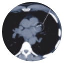
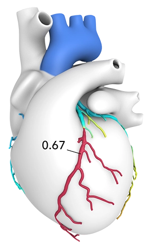
What is CT Cardiac Calcium Scoring?
One of our most accessible and valuable cardiac offerings is our low-cost, Low-Dose CT Cardiac Calcium Scoring exam.
Covered by most insurances, A CT Cardiac Calcium Scoring is a simple, non-invasive test that measures the amount of calcified plaque in your heart’s arteries. This specialized CT scan can help determine your risk of heart disease and heart attack.
It is important to note that a CT Cardiac Calcium Scoring exam is not diagnostic in itself. It is a screening exam and is typically used as a first line of detection and in combination with clinical evaluation by a cardiologist and other diagnostic tests, such as Coronary CTA (CCTA), to identify and evaluate the patient's risk of developing heart disease.
Benefits:
- Non-invasive
- Exam takes only a few seconds
- Low radiation
- Screening of the coronary arteries for calcified plaques
Who Should Consider CT Coronary Calcium Scoring:
- Men age 40-65
- Women age 50-70
- Patients at intermediate risk
When to consider starting to screen:
- Age 40 in men and 50 in women
- 10 years before age of 1st degree relative diagnosed with coronary artery disease (CAD)
Key Insights:
- Low to intermediate risk patients who score a "0" can often be managed with diet, exercise and sometimes medical therapy
- Does not detect non-calcified plaque
- Patients with prior coronary artery bypass grafts and stents or who have known CAD are not candidates for Calcium Scoring
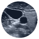
What is Carotid Ultrasound?
Carotid Ultrasound uses sound waves to produce pictures of the carotid arteries in the neck which carry blood from the heart to the brain. It is most frequently used to screen patients for blockage or narrowing of the carotid arteries which may increase the risk of stroke.
What is Ultrasound Echocardiogram (TTE)?
Ultrasound Transthoracic Echocardiogram is a diagnostic imaging test that uses high-frequency sound waves (Ultrasound) to create comprehensive images of the heart. The exam also includes Doppler Ultrasound, which measures the speed and direction of blood flow, helping to detect abnormal patterns and how well your heart is functioning.
Two of the most-common reasons (indications) for TTE:
- Suspected Heart Valve Disease: Issues such as mitral valve prolapse, aortic stenosis, or other valve abnormalities
- Unexplained Chest Pain or Shortness of Breath: Evaluates cardiac causes like heart failure or pericardial effusion
TTE helps your health care team find out:
- The size and shape of your heart, and the size, thickness and movement of your heart’s walls
- How your heart moves during heartbeats
- How well your heart is pumping blood
- If blood is leaking backwards through your heart valves (regurgitation)
- If the heart valves are too narrow (stenosis)
- If a tumor or infectious growth is around your heart valves
Key Insights:
- Low-cost, high-quality, and widely available tool
- Used for both initial diagnosis and ongoing management of cardiovascular disease
- If results are inconclusive or demonstrate abnormalities, a provider may refer for advanced imaging like Coronary CTA (CCTA) or Cardiac MRI
What is CCTA?
A heart imaging test that provides highly detailed images of the coronary arteries and anatomical data about the structure of the coronary arteries and the heart.
It helps to determine if plaque buildup has narrowed the coronary arteries (the blood vessels that supply the heart), which may cause chest pain or a heart attack. A referring provider may recommend a CCTA if calcified plaque has already been detected in the coronary arteries.
Benefits:
- Reliable alternative to invasive coronary angiography
- No radiation is left in your body after the scan is finished
- Provides very detailed images of many types of tissue
- Cost-effective for a wide range of medical problems
- Less sensitive to patient movement than MRI
- Implanted medical devices will not prevent the procedure
Who is CCTA for:
- Abnormal anatomy of the coronary arteries
- Low or intermediate risk for coronary artery disease
- Chest pain
- New or worsening symptoms with a previous normal stress test result
American Heart Association and the American College of Cardiology Risk Calculator

How CCTA with FFR CT works:
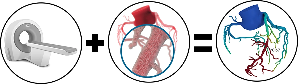
CT with Fractional Flow Reserve (FFR CT) is a physiologic simulation technique that models coronary flow from routine Coronary CT Angiography (CTA). At Lenox Hill Radiology, FFR CT is always interpreted in correlation with clinical and anatomic Coronary CTA findings.
FFR CT increases the specificity in the detection and evaluation of coronary artery disease, and decreases the prevalence of non-obstructive disease in invasive coronary angiography (ICA), which helps with decisions regarding revascularization vs. pharmacologic or other forms of treatment.
How can CCTA with FFR CT help:
- Increases specificity in the evaluation of coronary artery disease
- Reduces unnecessary exposure to radiation or complications from more invasive procedures
- Enables improved clinical decision-making
- Helps guide treatment for patients with CAD
Who is CCTA w/FFR CT for:
A physician may request the addition of an FFR-CT analysis to a CCTA, because of its ability to add functional information to the anatomical information provided by the CCTA.
Indications for performing FFR CT:
- Patients with 40-90% stenosis on a Coronary CTA in the setting of uncertain physiologic significance
Key Insights:
- Both anatomical and functional information provided allows for precise evaluation of the severity of CAD
- Can assist physicians with personalized treatment strategies
- Reduces likelihood for additional invasive follow-up studies
- Non-invasive
- According to a study published by the National Institute of Health (NIH), CCTA with added FFR CT “led to a reduction in (invasive) diagnostic testing and less revascularizations.”
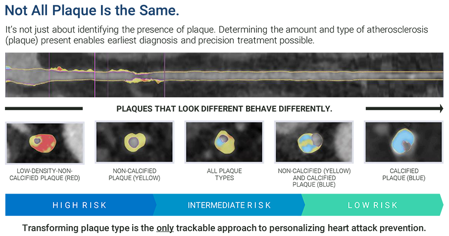
How CCTA with AI Plaque Analysis works:
AI Plaque Analysis uses machine learning software (artificial intelligence) to analyze images from a standard CCTA in order to quantify and characterize plaque in the arteries. The AI Plaque Analysis generates a 3D model of the coronary arteries, identifies their lumen and vessel walls, locates and quantifies stenoses, and identifies, quantifies and categorizes plaque. This additional process helps clinicians precisely identify and define atherosclerosis earlier.
Benefits:
- Post-processing analysis
- Used to guide treatment in symptomatic patients and for risk stratification
- Treatment response monitoring
- No additional scans needed - able to process from CCTA data
The artificial intelligence (AI) algorithms create a 2D model of the arterial lumen and outer wall. Included in the analysis are:
- Calcified, non-calcified and low-attenuation plaque areas
- Total plaque volume (TPV)
- For patients with acute or stable chest pain with 1-69% stenosis in their coronary arteries
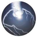
What is Cardiac MRI?
A Cardiac MRI, or cardiac magnetic resonance imaging, is a non-invasive test that uses a powerful magnetic field and radio waves to create detailed images of the structures within and around the heart. There are several types of cardiac MRI, all of which are used to evaluate how the heart, blood vessels and valves are working.
Lenox Hill Radiology’s subspecialized cardiac imaging radiologists are the New York metro region’s experts at the acquisition and interpretation of Cardiac MRI imaging. Lenox Hill Radiology uses the most advanced MRI acquisition techniques to ensure optimal data and the most accurate diagnosis.
Benefits:
- Lenox Hill Radiology's fleet of new MRI units are the region's most advanced
- No radiation
- Subspecialized cardiac imaging radiologists ensure accurate diagnosis
- Optimal for evaluating anatomy and function of the heart
Who Needs a Cardiac MRI:
- A cardiac MRI is usually ordered for a patient with more advanced or complex heart conditions
- Oncology patients on cardiotoxic medications
- Risk stratification before starting certain medications
- Optimal for evaluating anatomy and function of the heart
Please Note: LHR does NOT scan patients with pacemakers, defibrillators or other implanted electronic devices.
What is MUGA?
A multi-gated acquisition (MUGA) scan creates video images that show whether the lower chambers (ventricles) of the heart are pumping blood properly. MUGA uses intravenous material (radiotracers) to show how blood moves through the heart. MUGA can be used to check for pre-existing cardiac conditions prior to chemotherapy, or after treatment to assess possible side effects.
What is Nuclear Medicine Stress Test?
A nuclear medicine stress test, also known as a myocardial perfusion scan, is an imaging test that shows how blood flows to the heart at rest and during exercise. Using a small amount of radioactive material (radiotracer) administered by IV, the test measures blood flow through the heart’s arteries, and can help diagnose coronary artery disease (CAD).
Who is a candidate for a nuclear medicine stress test:
- Patients with suspected coronary artery disease
- Typically, patients who have chest pain suggestive of angina and intermediate to a high probability of acute coronary syndrome (ACS). However, it should not be performed in patients with ongoing or unstable conditions prior to the relief of symptoms.
- Patients with prior history of CAD with new or worsening symptoms
- Patients who have new-onset left bundle branch block (LBBB) or ventricular pacing in the EKG
- Patients with a recent ACS who were treated conservatively or partially revascularized within the past 3 months for evaluation of risk assessment
- Patients with a prior history of coronary artery bypass graft (CABG) of more than 5 years or percutaneous coronary intervention (PCI) of more than 2 years and are asymptomatic, one-time testing cardiac stress testing is appropriate
- Patients with dyspnea and suspected cardiac origin
- Patients with newly diagnosed congestive heart failure to evaluate reversible causes such as CAD
Nuclear Medicine Stress Test is only scheduled by the following:
Phone: (212) 599-5555
Email: LHROMICscheduling@RadNet.com