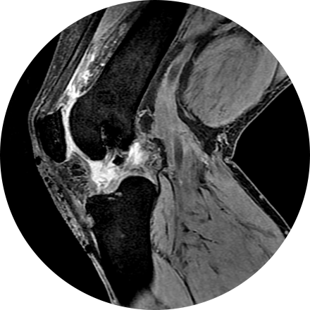
Musculoskeletal imaging addresses potential disorders related to a patient’s spine, bones, joints, muscles, soft tissues, ligaments and tendons. Evaluation of torn tendons, arthritis, cancer, systemic disease and post-traumatic injuries can all be made by musculoskeletal radiologists.
Some Musculoskeletal Imaging Procedures Include:
A dual-energy X-ray absorptiometry (DEXA) scan measures the density and mineral content in bone, most often in the hip or lower spine. It is the most accurate method of determining bone density and potential problems related to bone loss. This test is a valuable tool for diagnosing osteoporosis, which often has no symptoms until you suffer a fracture. A bone density scan can diagnose the disease at its earliest stages, which means you can begin receiving treatment to protect your bones sooner.
X-ray is the oldest and most economical form of medical imaging. During the procedure, radiation passes through the body onto “film” (now digitized and displayed on a computer screen). In neuroimaging, spinal X-rays are used to assess for the degree of spinal motion with flexion or extension.

